
MD-8500C HIGH FREQUENCY DIGITAL RADIOGRAPHY SYSTEM

MD-8500C HIGH FREQUENCY DIGITAL RADIOGRAPHY SYSTEM
Frequency: 260kHz
Tube current: 10-650mA
Tube voltage: 40~150kV
Imaging system: 17''x17''Flat Panel detector grey scale14bit Work station:1*24''LCD display
Product Usage
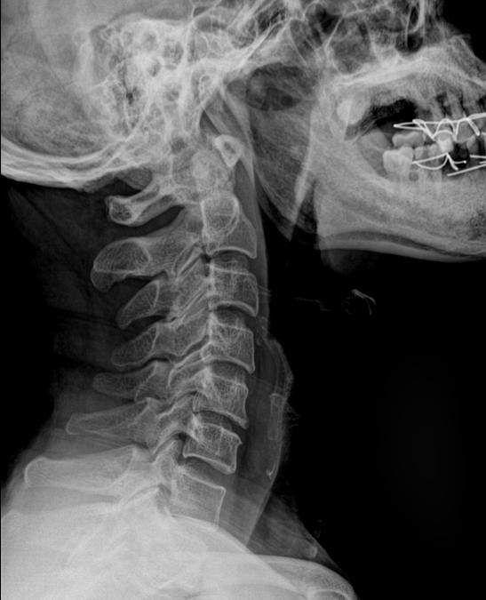
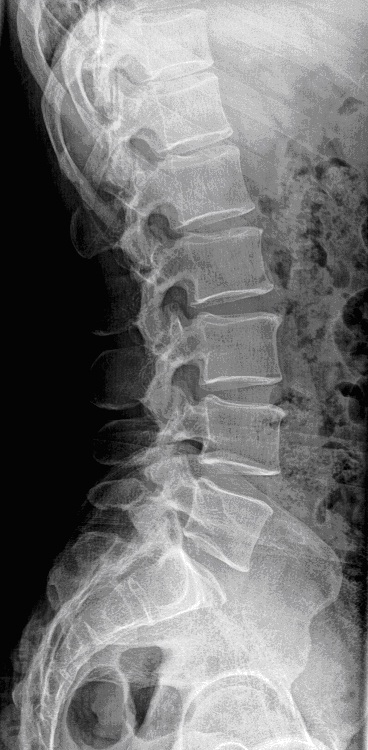
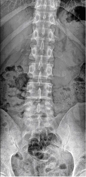
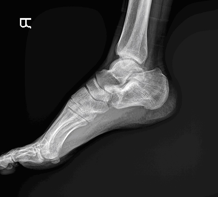
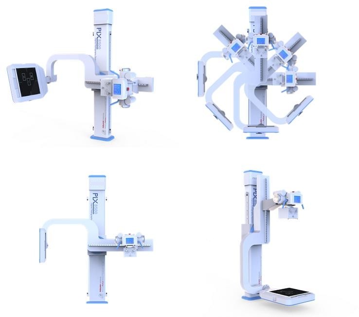
MD-8500C U-arm high frequency digital x-ray equipment can meet different parts radiography, such as head, chest, abdomen, waist, lumber, thoracic, pelvis, limbs etc. And meet different positions radiography, such as lay decubitus, normotopia, side position etc.
Optitionnal:diagnostic bed:F153

Product Specification
High frequency X-ray machine | Output power | 50kW | |
inverter frequency | 260kHz | ||
X-ray tube | Dual-focus X-ray tube | Small focus:0.6 Large focus:1.2 | |
Output power | 50 kW | ||
Anode Capacity | 111kJ(150KHU) | ||
Anode Angle | 12°
| ||
Speed of rotating anode | 3200rpm | ||
Tube Current | 10mA- 650mA
| ||
Tube voltage | 40-150kV | ||
mAs | 1-1000mAs
| ||
Exposure Time | 0.001-6.3s | ||
AEC | Option | ||
Digital Image System | Digital Detector | Panel size | 430mm*430mm
|
Pixel matrix | 3K*3K | ||
Spatial resolution | 3.7LP/mm | ||
Pixel size | 154um | ||
Output gray level | 16bit | ||
Imaging time | ≤9s | ||
DQE | ≥55% | ||
Acquisition module | Inside enhancement module | ||
Image Workstation
| Image information management
| Dicom image transmission Dicom film printing Dicom image storage | |
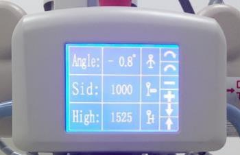
Mechanical structure & performance
| U-arm | Vertical movement range | ≥1250 mm(motorized control)
|
Focus-screen movement range | 1000mm-1800mm(motorized
| ||
Rotation range | -40°-+130°(motorized control)
| ||
Detector rotation | -45°-+45° | ||
X-ray Tube rotation | -180°-+180° | ||
Photography table (Optional) | Table size | 2000mm*650mm | |
Table height | ≤740mm | ||
Transverse movement | 200mm(electromagnetic lock) | ||
Longitudinal movement | 100mm(electromagnetic lock) | ||
Power supply | 380V 50/60Hz |
1: Type of generator and X-ray tube:
★ Advanced 260kHz high frequency high voltage type generator, realize 1ms instant exposure, high performance.
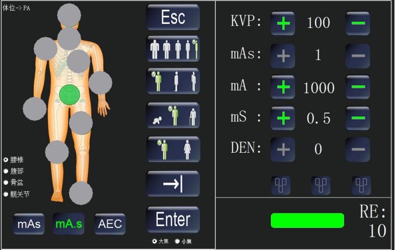
★ Three exposure method free change: KV, mAs two adjustment, KV, mA, s three adjustment and AEC function, to satisfy different habit of different doctors.
★ Rotate double anode 0.6 / 1.2, with high heat capacity of 150KHU
★ Digital micro-processed closed loop control and malfunction alarming system to reduce the dose of X-ray, protect the patients and doctors very well.
★ LCD touch screen, beautiful appearance and convenience to operate.
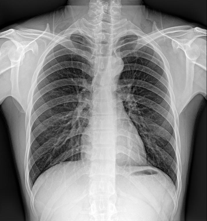
2: Flat Panel Detector
★ Apply with A-Si (Amorphous silicon) Toshiba imported Flat panel detector, which could give perfect digital images directly.
★ 3K×3K Acquisition matrix, 154um pixels size, and 3.7Lp/mm ultimate spatial resolution, with DQE values ≥ 55%
★ 17〞×17〞Large acquisition area and with the non-center processing technology, no matter the center and border, the quality of the image is the same.
★ The detector could be rotated ±45 degree along the axis direction, to satisfy the different photograph requirement of every body parts, such as Ankle joint, lateral spine
★ The detector has the self-protection function. It can stop to move when it detect the distance in front of the barrier.
3. Digital Working Station:
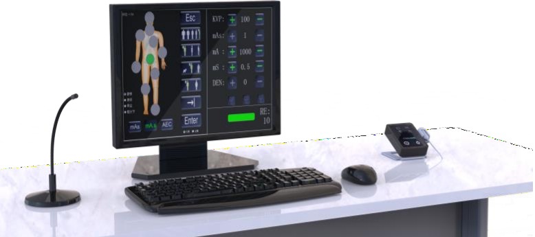
★ Case registration: Auto registration, be equipped with Dicom Worklist SCU. To simplify the input process for doctors, greatly reduce the amount of labour and greatly improve the working efficiency
★ Image Acquisition: Automatic window adjustment, Automatic cropping, Automatic transmit.
★ Image Processing: Tissue equilibrium, W/L adjustment, Gamma correction, interest district, reversed phase, noise reduction, smooth, sharpen, pseudo color, edge extraction, shadow compensation, filter nuclear, single window, dual-window, four windows, movement, right rotated 90°, left rotated 90°, level mirror image, vertical mirror image, magnifying glass, image zooming, reset, layer information, label character, drawing label, length measurement, angle measurement, rectangular length, rectangular area, elliptic length, elliptic area.
★ Dicom Image Transmit, Dicom Image Storage, Dicom Image viewing, Dicom Image printing.
★ Convenient to connect to the PACS system
4. Operation System:
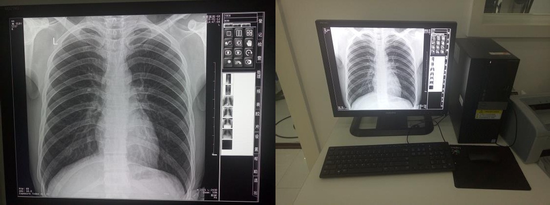 ★ Be equipped with 19〞imported LCD high resolution monitor screen,the delicate and richness degree of image is far higher than the normal medical monitor.
★ Brightness and Contrast are higher than 1000NIT, far higher than the normal LCD screen of 400 NIT.
★ These features can make the doctor diagnose more accurate and smooth.
★ Be equipped with the microphone and remote exposure control. The doctor can control outside the operating room.
★ Be equipped with various set of infrared facilities to protect the machine from the mis-operation of the doctors.
★ Optional PLXF153 operating room. Battery power supplied, Infrared unlock
★ Optional SONY, CODONICS film printer.
5. Mechanical Movement:
Film key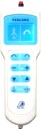
Hand Controller
★The self-designed and manufactured electric U-arm mainframe can move up and down, and rotate in a wide range, which can satisfy the requirements of multi-site photography.