
MD-6000 Dynamic FPD DRF
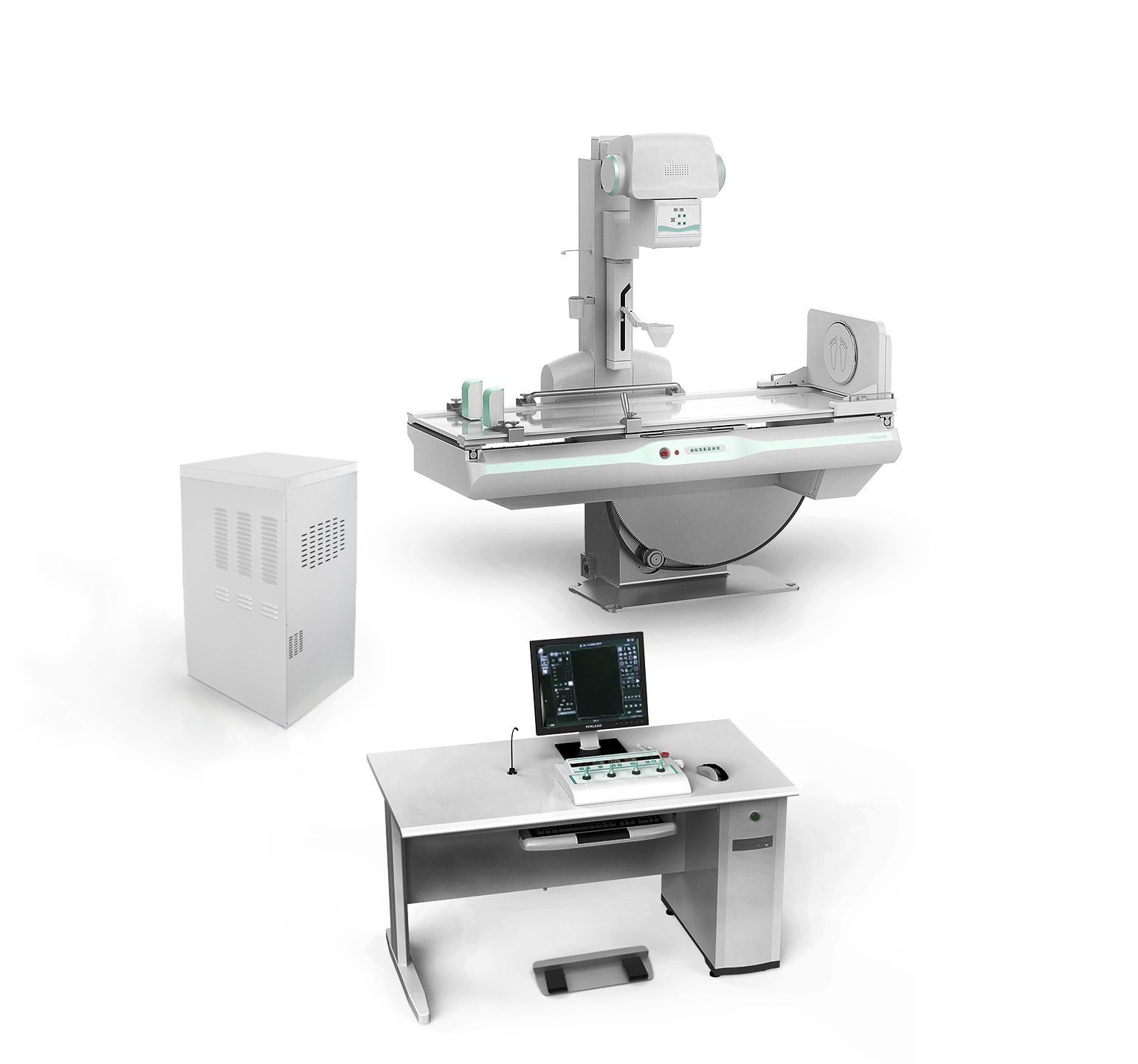
MD-6000 Dynamic FPD DRF
Configuration:
Item | Model | Quantity | Remark |
Console | KZT70A | 1 | MD- |
High voltage generator | FSQ65RF | 1 | MD- |
X ray tube | E7252X | 1 | Canon |
Collimator | XSQ20 | 1 | MD- |
Diagnostic table | ZDC20F | 1 | MD- |
Grid | 1.0/1.8m | 2 | JPI |
Dynamic flat panel detector | DRF-1717A | 1 | MD- |
Image acquisition workstation | DRA80 | 1 | MD- |
Image processing software(Optional) | DRA50 | 1 | MD- |
Accessory | MD-6000 | 1 | MD- |
Technical data
Item | Content | Technical parameter |
Power Supply | Voltage | 380V±38V |
frequency | 50Hz±1Hz | |
capacity | ||
internal resistance | ≤0.17Ω | |
High voltage generator (FSQ65RF) | Maximum output power | 65.5kW |
Main inverter frequency | 500khz, tolerance ± 20% | |
Radiography tube voltage | 40kV~150kV | |
Radiography tube current | 10mA~800mA regulation in steps | |
Radiography time | 1ms~ 10000ms regulation in steps | |
Radiography mAs | 0.1~1000 mAs | |
Fluoroscopy voltage | 40kV~ 125kV Continuously adjustable | |
Fluoroscopy current | 0.5mA~ 10mA (Continuously fluoroscopy) 5mA~ 20mA (pulse fluoroscopy) | |
Collimator(XSQ20) | Equivalent total filtration | |
X ray tube (E7252X) | Nominal anode input power | Big focus 75kW small focus 27kW |
Anode heat capacity | 210KJ(300kHU) | |
Component heat capacity | 900kJ(1250kHU) | |
Rotating anode speed | 9700rpm(180 Hz) | |
Tube focus | Big focus 1.2mm / Big focus 0.6mm | |
Target angle | 12° | |
Diagnostic (ZDC20F) | Table rotating range | +90 ° ~ 0 ° ~ -25 ° |
Longitudinal movement of point device | 1000mm±20mm | |
Lifting range of bed surface | Not less than 300mm; tolerance ±10mm | |
The load-bearing capacity of the bed: | 200kg | |
SID | 1000mm~1800mm;Tolerance ± 20 mm | |
Rotating foot pedal(optional) | 360°Infinite rotation. | |
Filter grid | 498.5x449mm 230L/INCH 10:1,focal length:100cm 498.5x449mm 103L/INCH 10:1,focal length:180cm | |
Dynamic flat panel detector (DRF-1717RF) | Effective area | 434mm(H)×434mm(V) |
Prime matrix | 3072(H)×3072(V) | |
Prime particle spacing | 139μm | |
Pulse fluoroscopy | 12fps / 1408 x 1408 16fps / 1024 x 1024 22fps / 768x768 | |
Continuous fluoroscopy | 13fps / 1408 x 1408 20fps / 1024 x 1024 30fps / 768x768 | |
Sequence radiography | 3fps/3072 x 3072 6fps/1536 x 1536 | |
Point piece | 3072 x 3072 1536 x 1536 | |
Spatial resolution | ≥3.7lp/mm | |
A / D Transformation | 16bit | |
Energy range | 40 ~ 150 kVp | |
Image output and control | Gigabit lan | |
Image acquisition workstation(DRA80) | Workstation software | Registration: routine registration, emergency registration, adding agreement, adding item, clearing information, starting inspection; Work list: list information, patient search to be examined, refresh to be examined list, delete examination, display column settings. Start inspection and emergency registration; Check list: list information, checked patient display and search, delete image, image storage, disc burning, add item, display column setting, modify check information; Patient's body type: thin adult, adult, fat adult; Photography parameter setting: exposure mode, frame rate setting, kVp, Ma, MS, MAS, AEC, focus selection; Perspective parameter setting: exposure mode, frame rate setting, kVp, Ma, ABS, time reset; Browsing tools: zoom, horizontal flip, vertical flip, left turn 90 degrees, right turn 90 degrees, zoom in, zoom out, original size, moving image, reverse color, adaptive size, ROI magnifying glass, magnifying glass, default window width and position, window width and position of interest area, visual window width and position, point gray value, advanced processing, elliptical gray measurement; System tools: text mark, front position mark, left mark, right mark, circle clipping, delete image, delete tool; Error reset, exposure indication, full screen, save current image, print Browsing tools: zoom, horizontal flip, vertical flip, left turn 90 degrees, right turn 90 degrees, zoom in, zoom out, original size, moving image, reverse color, adaptive size, ROI magnifying glass, magnifying glass, default window width and position, window width and position of interest area, visual window width and position, point gray value, advanced processing, elliptical gray measurement, image splicing; Measurement tools: arrow, cardiothoracic ratio (CTR), distance measurement, angle measurement, spine measurement; System tools: text mark, front position mark, left mark, right mark, circle clipping, delete image, delete tool; Report, save current image, print Report editing: patient information display and editing, photo image selection, report content template selection, report description, report conclusion, report description + conclusion, edit knowledge base, report doctor, audit doctor, report time, print template, setting and saving report; Report printing: fast printing, printing report Image archiving, burning, printing: delete image, image storage, browse image, report, lock / unlock, storage queue, print queue; Disc recording: volume label, save settings, file compression, file structure; Printing: DICOM printer, local printer System settings: system, annotation information, tools, others; Hardware configuration: syncbox, high voltage, detector, collimator, DAP; Network configuration: local, worklist, netstore, local store, printing; Inspection management: basic information, positioning information, hardware parameters, image processing, inspection protocol;
Quality management: search, system management;
User management: add, update, delete, permission.
|
Image processing software(Optional) | Case inquiry | 1) Support patient medical status control, including; unread, read, reported, audited, etc.; 2) Support multiple query methods such as detailed and fuzzy; 3) Support single item and multiple condition combination query; 4) Support patient information query, such as Chinese name, visit number, examination number, outpatient, hospitalization number, etc.; 5) Support inspection information query, such as inspection date, inspection location, inspection equipment, etc.; 6) Support diagnosis conclusions, keyword query. 7) Support to increase and modify patient information. |
Image processing software(Optional) | Report processing | 1) A variety of report printing formats can be provided for different image inspections; 2) Support multiple graphic report formats 3) Support report preview and printing 4) The report printing authority can be set; 5) Support choose different printers in the network to output reports; 6) The printed report is marked; 7) When opening or editing, it can automatically read and display related images in the image display window; 8) Support the search of patients' previous reports; 9) Support the import function of the patient's previous reports; 10) Provide a library of expert report templates corresponding to various image inspections; 11) Support quick calling of templates; 12) Users can freely edit, modify, delete and add modules; 13) Support the import of report templates; |
Image processing software(Optional) | Image processing | 1) Support single-screen and multi-screen display of images; 2) Support various color monitors and high-resolution black and white medical professional monitors; 3) Supports display of image index in two ways: sequence and image tile; 4) Supports quick navigation of patients, examinations, and sequences through ton thumbnails; 5) Support user-defined display layout; 6) Support to restore the original image function; 7) Multi-frame image dynamic playback display and tile display; 8) Support mouse wheel, keyboard to quickly flip through images; 9) Supports easy adjustment of interactive window width and window position with the movement of the mouse; 10) Support multi-window adjustment of the same image on the same screen; 11) The window width and window level value can be preset according to the imaging equipment and the filming position; 12) Support anti-window function; 13) Support image automatically adapt to window display; 14) Support image movement; 15) Support stepless reduction, enlargement, and partial magnifying glass; 16) Support clockwise, counterclockwise rotation, horizontal flip, vertical flip; 17) Support text annotation, graphics, arrow mark; 18) Support users to add, delete, edit, move, and an annotation on the image arbitrarily; 19) The user can show or hide the annotations on the image; 20) Support distance measurement, angle measurement, ellipse measurement; 21) Support the measurement of cardiothoracic ratio; 22) Support the measurement of CT value and positioning line; 23) Support image JPEG and other formats output; |
Image processing software(Optional) | Film printing process | 1) Support networking with DICOM cameras; 2) Support DICOM standard film printing related services; 3) Support flexible image printing and typesetting; 4) Support image horizontal and vertical printing typesetting; 5) Image related information can be on the corresponding film at the same time; 6) Provide a variety of printing parameter settings.
|
Image processing software(Optional) | Image sending function | Send the image to other DICOM receiver devices |
Image processing software(Optional) | Burn disc function | Export selected exams to CD/DCD discs, with CD player software |
Image processing software(Optional) | Image data archiving | 1) Support all kinds of medical imaging equipment with DICOM interface, including CT, MR, CR, DR, DSA, ECT, PET/CT, digital breast machine, digital gastrointestinal machine, etc.; 2) Support to obtain the image data of DICOM standard output device and store it in DICOM format; 3) Supports importing DICOM images and deleting data. 4) Support WORKLIST query. 5) Complete log records, through the log files, you can know the operating status of the system technically, and troubleshoot early. |