
MD-8000C Dynamic FPD DRF
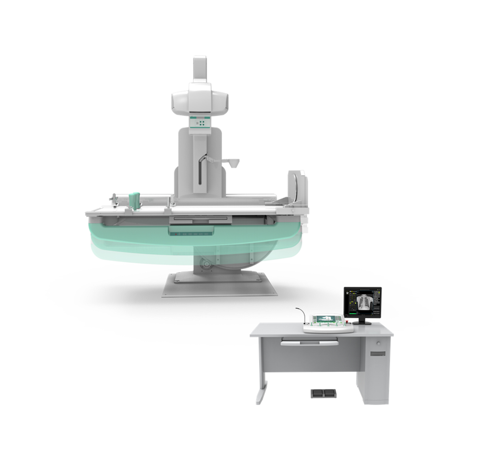
MD-8000C Dynamic FPD DRF
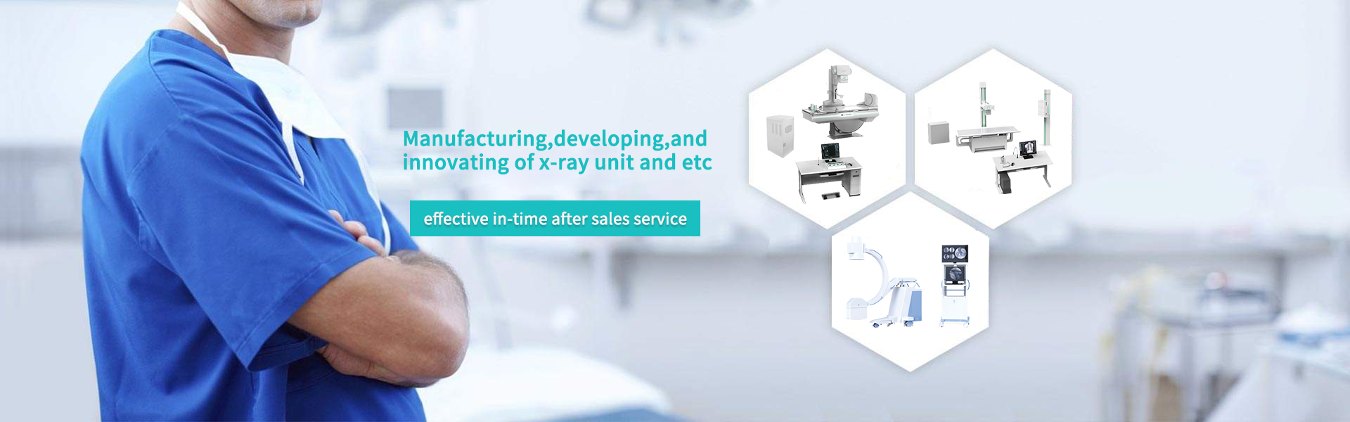
Product Use:
This machine is equipped with digital fluoroscopy, digital spot photography and digital DR photography. It can be used for chest and abdominal fluoroscopy, gastrointestinal angiography, urogenital angiography, cholangiography and lower extremity veins. It can be used for head and chest. DR photography of the abdomen and limbs; can also be used for fracture reconstruction under fluoroscopy, foreign bodies, etc.; can be used for peripheral angiography and interventional therapy.
Configuration:
Item | Model | Quantity | Remark |
Console | KZT70 | 1 | MD- |
High voltage generator | FSQ65 | 1 | MD- |
X ray tube | E7254FX | 1 | Canon |
Collimator | XSQ40 | 1 | MD- |
Diagnostic table | ZDC50 | 1 | MD- |
Grid | 1.0/1.8m | 2 | JPI |
AEC | SSMC601 | 1 | Clemmond |
DAP | d147 | 1 | MD- |
Dynamic flat panel detector | DRF-1717RF | 1 | MD- |
Image acquisition workstation | DRA80 | 1 | MD- |
Stitching function | optional | 1 | MD- |
Accessory | MD-8000C | 1 | MD- |
Technical data:
Item | Content | Technical parameter |
Power Supply | Voltage | 380V±38V |
frequency | 50Hz±1Hz | |
capacity | ||
internal resistance | ≤0.11Ω | |
High voltage generator (FSQ65) | Maximum output power | 65.5kW |
Main inverter frequency | 440khz, tolerance ± 10% | |
Radiography tube voltage | 40kV~150kV | |
Radiography tube current | 10mA~800mA regulation in steps | |
Radiography time | 1ms~ 10000ms regulation in steps | |
Radiography mAs | 0.1~1000 mAs | |
Fluoroscopy voltage | 40kV~ 125kV Continuously adjustable | |
Fluoroscopy current | 0.5mA~ 10mA (Continuously fluoroscopy) 5mA~ 20mA (pulse fluoroscopy) | |
Collimator(XSQ40) | Equivalent total filtration | |
X ray tube (E7254FX) | Nominal anode input power | Big focus 102kW small focus 40kW |
Anode heat capacity | 285KJ(400kHU) | |
Component heat capacity | 950kJ(1339kHU) | |
Rotating anode speed | 9700rpm(180 Hz) | |
Tube focus | Big focus 1.2mm / Big focus 0.6mm | |
Target angle | 12° | |
Diagnostic (ZDC50) | Table rotating range | +90 ° ~ 0 ° ~ -25 ° |
Longitudinal movement of point device | 900mm | |
Table Lifting | 300mm | |
Lateral movement range of table surface | 220mm | |
SID | 800mm~1800mm | |
Filter grid | 18 "× 18" 215lp1 12:1 100cm and 180cm filter grids, one for each, manual switching | |
Dynamic flat panel detector (DRF-1717RF) | Effective area | 434mm(H)×434mm(V) |
Prime matrix | 2816(H)×2816(V) | |
Prime particle spacing | 154μm | |
Pulse fluoroscopy | 12fps / 1408 x 1408 16fps / 1024 x 1024 22fps / 768x768 | |
Continuous fluoroscopy | 13fps / 1408 x 1408 20fps / 1024 x 1024 30fps / 768x768 | |
Spatial resolution | ≥3.7lp/mm | |
A / D Transformation | 16bit | |
Energy range | 40 ~ 150 kVp | |
Working mode | 8 | |
Image output and control | Gigabit lan | |
Image acquisition workstation(DRA80) | Workstation software | Registration: routine registration, emergency registration, adding agreement, adding item, clearing information, starting inspection; Work list: list information, patient search to be examined, refresh to be examined list, delete examination, display column settings. Start inspection and emergency registration; Check list: list information, checked patient display and search, delete image, image storage, disc burning, add item, display column setting, modify check information; Patient's body type: thin adult, adult, fat adult; Photography parameter setting: exposure mode, frame rate setting, kVp, Ma, MS, MAS, AEC, focus selection; Perspective parameter setting: exposure mode, frame rate setting, kVp, Ma, ABS, time reset; Browsing tools: zoom, horizontal flip, vertical flip, left turn 90 degrees, right turn 90 degrees, zoom in, zoom out, original size, moving image, reverse color, adaptive size, ROI magnifying glass, magnifying glass, default window width and position, window width and position of interest area, visual window width and position, point gray value, advanced processing, elliptical gray measurement; System tools: text mark, front position mark, left mark, right mark, circle clipping, delete image, delete tool; Error reset, exposure indication, full screen, save current image, print Browsing tools: zoom, horizontal flip, vertical flip, left turn 90 degrees, right turn 90 degrees, zoom in, zoom out, original size, moving image, reverse color, adaptive size, ROI magnifying glass, magnifying glass, default window width and position, window width and position of interest area, visual window width and position, point gray value, advanced processing, elliptical gray measurement, image splicing; Measurement tools: arrow, cardiothoracic ratio (CTR), distance measurement, angle measurement, spine measurement; System tools: text mark, front position mark, left mark, right mark, circle clipping, delete image, delete tool; Report, save current image, print Report editing: patient information display and editing, photo image selection, report content template selection, report description, report conclusion, report description + conclusion, edit knowledge base, report doctor, audit doctor, report time, print template, setting and saving report; Report printing: fast printing, printing report Image archiving, burning, printing: delete image, image storage, browse image, report, lock / unlock, storage queue, print queue; Disc recording: volume label, save settings, file compression, file structure; Printing: DICOM printer, local printer System settings: system, annotation information, tools, others; Hardware configuration: syncbox, high voltage, detector, collimator, DAP; Network configuration: local, worklist, netstore, local store, printing; Inspection management: basic information, positioning information, hardware parameters, image processing, inspection protocol;
Quality management: search, system management;
User management: add, update, delete, permission.
|